
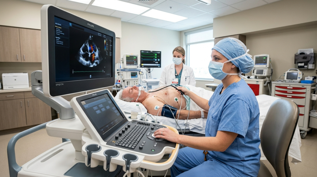
A TEE test, or transesophageal echocardiography, is a specialized form of cardiac ultrasound that produces detailed images of the heart from inside the esophagus. Instead of placing an ultrasound transducer on the chest, clinicians guide a flexible probe through the mouth and into the esophagus, which lies close to the heart.
This position creates an alternative acoustic window and can make certain cardiac structures easier to visualize than with a conventional transthoracic echocardiogram (TTE). TEE does not use ionizing radiation; it uses high-frequency sound waves to generate moving images of cardiac anatomy and blood flow.
TEE is not simply a more advanced replacement for every standard echocardiogram. Rather, it is a specialized imaging technique used when a particular clinical question requires detailed views, when transthoracic imaging is inadequate, or when real-time cardiac imaging is useful during selected procedures.
What Is a TEE Test?
What does TEE stand for?
TEE stands for transesophageal echocardiography.
The term can be understood in two parts:
- Transesophageal means the ultrasound probe is positioned through the esophagus.
- Echocardiography refers to imaging the heart using ultrasound waves.
The TEE probe contains an ultrasound transducer. It sends sound waves into the surrounding tissues and receives the returning echoes. A computer processes these signals to create images of the heart and nearby structures in real time.
Because ultrasound does not rely on X-rays, a TEE examination does not expose the patient to ionizing radiation.
How is TEE different from a standard echocardiogram?
The conventional approach is transthoracic echocardiography (TTE). During TTE, a transducer is moved over the chest wall to obtain images of the heart.
TEE uses a flexible probe positioned inside the esophagus. Because the esophagus runs immediately behind the heart, the transducer can be placed much closer to several cardiac structures. This can provide useful views when the chest-wall approach is limited by anatomy, lung tissue, body habitus, previous surgery, or other factors.
The two techniques are therefore best viewed as complementary rather than competing technologies. Professional echocardiography guidance describes TEE as an imaging method that can provide additional information for selected diagnoses and cardiac interventions.
How Transesophageal Echocardiography Produces Heart Images
The role of the transducer
At the end of the TEE probe is an ultrasound transducer. The transducer converts electrical energy into high-frequency sound waves and then detects the echoes returning from tissues.
When ultrasound encounters boundaries between tissues with different acoustic properties, part of the sound energy is reflected back toward the transducer. The system uses the returning signals to determine where structures are located and how they appear.
Modern TEE systems can use sophisticated transducer designs and imaging technologies, including multiplane and three-dimensional capabilities. The American Society of Echocardiography notes that advances in flexible phased-array and matrix-array transducers have expanded the imaging capabilities of TEE.
From ultrasound echoes to cardiac images
The ultrasound system analyzes the timing and characteristics of returning echoes to construct an image.
One important factor is depth. The system can estimate how far a reflecting structure is from the transducer based on how long the ultrasound signal takes to return.
Image quality also depends on factors such as resolution, acoustic windows, probe position, transducer characteristics, tissue interfaces, and operator technique. Echocardiographic images can show moving structures, allowing clinicians to observe valve motion, chamber activity and blood-flow patterns rather than relying on static anatomy alone.
Doppler techniques add another layer of information by evaluating movement of blood relative to the ultrasound beam. Color Doppler and spectral Doppler can therefore contribute functional information alongside conventional two-dimensional imaging.
Why positioning the probe in the esophagus matters
The esophagus lies behind the heart, separated from many cardiac structures by relatively little tissue. Placing the ultrasound transducer in this location can reduce some of the obstacles encountered when imaging through the chest.
This anatomical relationship is particularly useful for examining structures such as the left atrium, mitral valve, atrial septum, portions of the aorta and the left atrial appendage.
TEE guidelines from the American Society of Echocardiography describe standardized imaging views while also emphasizing that the examination may need to be adapted to the patient’s anatomy, pathology and clinical question.
What Can a TEE Examination Show?
The exact images obtained during a TEE depend on why the examination is being performed. A comprehensive examination may involve multiple standardized views, while a focused study may concentrate on a specific structure or clinical question.
Heart valves
TEE can provide detailed views of cardiac valves and their movement. This can be important when clinicians need to assess valve anatomy, prosthetic valves, suspected valve infection or abnormalities associated with valve function.
For selected structural-heart procedures, TEE can also contribute to pre-procedure anatomical assessment and procedural planning.
Chambers, septa and selected cardiac structures
TEE can visualize the atria and ventricles as well as structures such as the interatrial septum. The closer position of the transducer can be particularly useful when an external echocardiographic window does not provide sufficient detail.
The technique can also be used to examine portions of the great vessels and thoracic aorta, depending on the clinical indication and imaging protocol.
🏢 Featured Medical Devices Directory Listing
Looking for verified and trusted medical devices partners related to this topic? Explore our comprehensive directory.
Explore Directory →Blood flow and Doppler assessment
Echocardiography is not limited to anatomical imaging. Doppler methods allow assessment of blood movement through cardiac chambers and across valves.
This information can help clinicians evaluate patterns such as abnormal or turbulent flow and contribute to assessment of valve function and other hemodynamic questions.
The images and measurements are interpreted in their clinical context by appropriately trained professionals rather than being treated as standalone measurements.
The left atrial appendage and intracardiac abnormalities
The left atrial appendage (LAA) is a small structure connected to the left atrium that can be important in selected evaluations for intracardiac thrombus.
TEE is commonly used when detailed assessment of the LAA or other potential cardiac sources of embolic material is clinically relevant. However, the purpose of an examination varies from patient to patient; not every TEE is performed to look for a thrombus.
Why Might a Clinician Recommend a TEE?
TEE is generally selected because it can answer a specific imaging question that may not be adequately addressed by TTE alone.
When standard echocardiography does not provide enough detail
A TTE examination can sometimes be technically limited. If the chest-wall acoustic window does not adequately show an important structure, TEE may provide another way to obtain the necessary images.
This complementary relationship is one of the central reasons TEE remains important in cardiovascular imaging.
Evaluating suspected valve or structural heart abnormalities
TEE can be useful when detailed anatomical information is required for selected valve disorders, suspected infective endocarditis, prosthetic valve assessment and structural heart abnormalities.
The decision depends on the clinical question and the information already available from other examinations.
Looking for certain intracardiac sources of embolic events
In selected patients undergoing evaluation for stroke, transient ischemic attack or other embolic events, TEE may be used to examine cardiac structures that could provide a potential source of embolic material.
This is one reason the technique has an important role in cardiovascular diagnostic imaging, although the appropriate investigation depends on the individual’s clinical circumstances.
Before or during selected cardiac procedures
TEE is also used in selected cardiac surgeries and catheter-based interventions.
During structural-heart procedures, real-time echocardiographic imaging can help clinicians understand anatomy, guide procedural steps and assess results. ASE guidance specifically describes the role of TEE in structural-heart intervention planning and imaging.
This broader use illustrates how modern diagnostic technologies increasingly overlap with procedural medicine. Similar developments are occurring throughout the medical diagnostics field, where advances in diagnostic testing are increasingly connected with clinical decision-making and treatment workflows. advances in diagnostic testing
What Happens During a TEE Procedure?
A TEE examination generally follows several stages, although exact protocols differ between hospitals and clinical settings.
Before the probe is inserted
Before the examination, the healthcare team typically reviews relevant medical history, medications, allergies and conditions that could affect probe insertion or sedation.
Patients should tell the clinical team about swallowing difficulties, known esophageal disorders, previous esophageal procedures and other relevant medical conditions. Potential contraindications and risk factors need to be evaluated rather than assumed to be the same for every patient.
Fasting is also commonly required before TEE, particularly because sedation may be used and because the probe passes through the mouth and esophagus. The exact fasting instructions should come from the facility performing the examination.
Positioning and throat preparation
The patient is generally positioned on their side or in another position appropriate for the examination. Monitoring equipment is used to observe parameters such as heart rhythm and oxygen levels.
A local anesthetic may be applied to the throat to reduce discomfort and help control the gag reflex. Sedative medication may also be used depending on the clinical setting and circumstances.
Passing the TEE probe
Once preparation is complete, the flexible ultrasound probe is guided through the mouth and into the esophagus. The patient may be asked to swallow as the probe is advanced.
The probe is then positioned and manipulated to obtain different views of the heart. Because modern TEE probes can acquire multiple imaging planes, the operator can examine cardiac structures from different orientations.
Capturing and analyzing the images
The transducer emits ultrasound and records returning echoes while the operator adjusts the probe and imaging controls.
Depending on the examination, the team may obtain two-dimensional images, Doppler measurements and, where appropriate, three-dimensional views. ASE’s comprehensive TEE guidance describes a standardized set of views while recognizing that the final examination should be adapted to the clinical situation.
What happens after the examination?
After the probe is removed, patients may be observed while the effects of sedation wear off if sedation was used.
Temporary throat soreness or a scratchy voice can occur. If the throat remains numb from local anesthetic, patients may be instructed to wait before eating or drinking normally.
Post-procedure instructions vary by facility and should be followed carefully.
🏢 Featured Medical Devices Directory Listing
Looking for verified and trusted medical devices partners related to this topic? Explore our comprehensive directory.
Explore Directory →How to Prepare for a TEE Test
Fasting before TEE
Patients are often instructed not to eat or drink for several hours before a TEE. The reason is related to procedural safety and the use of sedation or anesthesia in many settings.
There is no single fasting schedule that should be applied universally. The performing facility should provide the exact instructions.
Medication and medical-history considerations
The clinical team should know about prescription medicines, over-the-counter medicines, allergies and relevant medical conditions.
In particular, swallowing difficulties and known esophageal problems can be important because the procedure involves passing a probe through the esophagus. Published safety literature identifies several esophageal and upper gastrointestinal conditions that may require careful assessment before TEE.
Patients should not independently stop or change prescribed medication solely because a TEE is scheduled. Medication instructions should come from the healthcare team responsible for the procedure.
Arranging transportation when sedation is used
If sedative medication is administered, patients may not be able to drive afterward. Some facilities therefore require or strongly recommend that patients arrange transportation home.
Requirements vary according to the sedation protocol and facility, so these arrangements should be confirmed before the appointment.
TEE vs. TTE: What Is the Difference?
| Feature | TEE | TTE |
| Transducer location | Inside the esophagus | Outside the chest |
| Invasiveness | Semi-invasive/internal probe | Non-invasive |
| Acoustic window | Close to the heart from the esophagus | Through the chest wall |
| Sedation/anesthesia | May be used depending on circumstances | Generally not required |
| Typical role | Detailed assessment of selected structures and procedures | Common external cardiac ultrasound examination |
Why TEE can provide different views
The principal technical advantage of TEE is the location of the transducer. From inside the esophagus, the probe can obtain views of structures that may be difficult to image through the chest wall.
This can be particularly useful for posterior cardiac structures, valves, the left atrial appendage and selected portions of the aorta.
Why TTE remains important
TTE remains a central cardiac imaging technique because it is non-invasive, generally accessible and does not require passage of a probe into the esophagus.
For many routine cardiac questions, TTE can provide the necessary information without the additional preparation and procedural considerations associated with TEE.
The choice between TTE and TEE therefore depends on the anatomical or clinical question rather than a simple hierarchy in which one technique is always preferable.
What Are the Risks and Limitations of TEE?
TEE is generally considered a safe procedure when performed by appropriately trained professionals, but it is not risk-free.
Common temporary effects
Temporary throat soreness, difficulty swallowing immediately after the examination, or a scratchy voice may occur. Sedative medicines can also produce temporary effects such as drowsiness or nausea.
Less common but clinically important complications
Because the probe passes through the mouth and esophagus, mechanical injury is possible. Potential complications include injury to the teeth, gums, throat or esophagus, bleeding, abnormal heart rhythm and, rarely, esophageal perforation.
The published literature describes serious esophageal complications as uncommon, although risk can vary according to patient characteristics and underlying esophageal pathology.
Sedation or anesthesia also introduces its own potential risks.
Why certain patients may require additional assessment before TEE
Known esophageal strictures, certain diverticula, previous esophageal surgery, perforation and other gastrointestinal or anatomical conditions may affect the safety of probe insertion.
Importantly, published guidance does not support treating every possible risk factor in exactly the same way. Clinical assessment is required to determine whether TEE is appropriate and how it should be performed.
How Long Does a TEE Test Take?
There is no single duration that applies to every TEE examination.
The actual imaging portion can be relatively brief, but the total appointment includes preparation, throat anesthesia, possible sedation, image acquisition and recovery.
For example, Mayo Clinic Healthcare notes that the examination itself may take around 20–30 minutes in its setting, while the overall visit can be substantially longer because of preparation and recovery. Other clinical situations, particularly complex procedural or intraoperative examinations, can require different workflows.
Why procedure duration can vary
The duration may depend on:
- The number and type of images required
- The complexity of the clinical question
- Whether Doppler or three-dimensional imaging is needed
- Whether TEE is being used during a cardiac procedure
- The patient’s anatomy and ability to tolerate the examination
- Preparation and recovery requirements
How Are TEE Results Interpreted?
What the echocardiography team evaluates
Depending on the indication, a TEE examination may evaluate:
🏢 Featured Medical Devices Directory Listing
Looking for verified and trusted medical devices partners related to this topic? Explore our comprehensive directory.
Explore Directory →- Cardiac anatomy
- Heart valves and valve motion
- Cardiac chambers
- Septal structures
- Blood-flow patterns
- Left atrial appendage
- Selected portions of the aorta and great vessels
- Findings relevant to a planned cardiac procedure
Not every examination evaluates every structure in equal detail. ASE guidance emphasizes that imaging protocols can be tailored to the clinical question and the patient’s anatomy.
How the findings become a clinical report
The acquired images and measurements are reviewed by qualified professionals and incorporated into an echocardiography report.
Interpretation does not normally depend on one isolated image or measurement. Findings are considered alongside symptoms, medical history, physical examination and other diagnostic information.
This distinction is important because an imaging finding by itself does not necessarily establish a diagnosis or determine treatment.
ASE’s updated reporting guidance also emphasizes standardized terminology and reporting practices across echocardiography, including TEE, to improve communication among clinical teams and healthcare systems.
What Makes TEE Particularly Valuable in Modern Cardiac Imaging?
High-detail imaging close to cardiac structures
TEE combines the physical advantage of placing the transducer close to the heart with sophisticated ultrasound imaging.
The result is an imaging approach that can provide detailed views of anatomy that may be challenging to visualize externally. Advances in probe design, multiplane imaging and three-dimensional echocardiography have expanded what can be assessed using TEE.
Role in image-guided cardiac procedures
The value of TEE extends beyond diagnosis. Real-time imaging can help clinicians plan and monitor selected cardiac and structural-heart procedures.
For structural interventions, TEE may contribute information about anatomy, disease severity and device suitability. During selected procedures, imaging can also provide immediate feedback about cardiac structures and procedural results.
The importance of operator expertise and imaging equipment
A TEE examination is not simply a matter of inserting a sophisticated probe and pressing a button.
Image quality depends on the ultrasound platform, transducer capabilities, probe position, imaging settings, patient anatomy and the skill of the person acquiring and interpreting the images.
This is particularly relevant to the medical-device ecosystem. Improvements in ultrasound transducers, imaging software, three-dimensional reconstruction and reporting systems can expand diagnostic capabilities, but the technology still depends on appropriate clinical use and expert interpretation.
Key Questions to Ask Before a TEE Examination
A patient or healthcare learner may find it useful to understand the main questions surrounding the examination.
Questions about preparation
- How long should fasting continue?
- Can regular medications be taken before the examination?
- Will sedation be used?
- Is someone required to accompany the patient home?
Questions about the examination
- Why is TEE being recommended?
- What cardiac structure or clinical question is being evaluated?
- How will the images be interpreted?
- When and how will the results be communicated?
Questions about individual risks
- Are there swallowing or esophageal conditions that should be discussed before the procedure?
- Are there particular risks associated with the individual’s medical history?
- What post-procedure symptoms should prompt contact with the healthcare team?
These questions do not replace medical advice, but they can help patients understand the purpose and practical requirements of the examination.
Conclusion
A TEE test is a specialized ultrasound-based cardiac imaging procedure that places the transducer in the esophagus to obtain detailed views of the heart and selected nearby structures. Its value comes from the combination of anatomical positioning, ultrasound technology, real-time imaging and professional interpretation.
TEE can be useful when transthoracic echocardiography does not provide enough information, when detailed assessment of selected cardiac structures is required, or when real-time imaging is needed during certain cardiac procedures. At the same time, TEE involves a probe passing through the esophagus and therefore has different preparation requirements and risks from a standard TTE examination.
For medical-device and diagnostic-technology professionals, TEE also illustrates how imaging hardware, transducer engineering, software, operator expertise and clinical workflows work together to produce actionable cardiovascular information. Rather than replacing routine echocardiography, TEE occupies a specialized role within the broader cardiac imaging ecosystem.